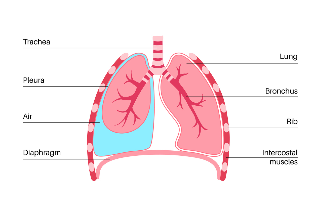
Anatomy of the Pleura
The pleura consists of two layers:
- Visceral pleura (Pleura visceralis): The visceral pleura is the inner layer of the pleura and envelops both lungs. At the lung hilum (entry point of the pulmonary arteries, pulmonary veins, and bronchi into the lung), the visceral pleura transitions into the outer layer of the pleura (parietal pleura).
- Parietal pleura (Pleura parietalis): The parietal pleura is the outer layer of the pleura and lines the chest wall (thoracic wall) and the upper surface of the diaphragm. Likely only the parietal pleura is innervated and is responsible for pain, for example in pleuritis.
Between the visceral pleura and the parietal pleura exists a sliding layer, because a pleural space (Cavitas pleuralis) is present. This space is filled with a few milliliters of serous fluid, the pleural fluid.
At the outer margins of the diaphragm and in the region of the mediastinum, the pleura forms so-called recesses (outpouchings), which serve as reserve folds during inspiration to allow expansion of the lung. A total of 4 recesses are distinguished:
- Recessus costodiaphragmaticus
- Recessus costomediastinalis
- Recessus phrenicomediastinalis
- Recessus vertebromediastinalis
The region of the pleura that extends superiorly (cranially) beyond the first rib is called the pleural cupula (Cupula pleurae).
Physiology of the Pleura:
The pleura serves as a sliding space between the chest wall and the lung. Through negative pressure in the pleural space (−5 cm H2O) and the capillary adhesion of the two pleural layers, the lung follows the movements of the chest wall and diaphragm, so that the musculature of these structures enables respiratory movements.
Pathophysiology of the Pleura:
When the negative pressure in the pleural space is eliminated by an injury (for example, pneumothorax), the lung can no longer follow the excursions of the respiratory muscles. With further pressure increase, collapse of the affected lung occurs.
In the pleural recesses, an abnormal amount of fluid (blood, exudate, pus, etc.) can accumulate. This is then referred to as pleural effusion (or hemothorax, pleural empyema).
Diseases of the Pleura
- Accumulation of air in the pleural space: pneumothorax; occurring spontaneously or due to a traumatic event
- catamenial pneumothorax in endometriosis;
- Accumulation of air in the pleural space under pressure: tension pneumothorax;
- Accumulation of blood in the pleural space: hemothorax; occurring after trauma or in pleural tumors;
- Accumulation of fluid or pus in the pleural space in pleuritis;
- Inflammation of the pleura (pleuritis); also in tuberculosis (TB);
- Tumors of the pleura; pleural carcinomatosis (pleural metastases from another primary tumor); primary pleural tumors, especially malignant pleural mesothelioma (MPM). Benign solitary pleural fibroma.