Thorax Schweiz
  • Home
  • Anatomy & Disease Patterns
    • Thorax: Function and Anatomy
    • The Airways (Trachea & Bronchi)
    • The Lungs (Pulmo)
    • The Pleura
    • The diaphragm (diaphragma)
    • The mediastinum
    • The Chest (Thorax)
    • The Esophagus
  • Patients & Relatives
    • Patients & Relatives
    • Support Groups and Organizations
    • Your General Practitioner’s Office
  • Prevention
    • Preventive Healthcare
    • The Importance of Prevention
    • Smoke-Free Living
    • Follow-up care
    • Rehabilitation
  • Podcast
  • Blog
  • About Us
    • About Thorax Switzerland
    • The medical team at Thorax Schweiz
    • Additional Experts
    • THE INITIATORS OF THORAX SWITZERLAND
  • Contact
  • English
    • Français (French)
    • Deutsch (German)
    • Italiano (Italian)
Author

csadmin

csadmin

Thorax: Function and Anatomy

The mediastinum

by csadmin 14. January 2024
written by csadmin
Anatomy of the mediastinum

In clinical practice, the mediastinum is commonly divided into an anterior, middle, and posterior compartment.

The anterior compartment (mediastinum anterius) comprises the space between the sternum and the pericardium, as well as the costomediastinal recess of the pleural sacs. The contents of this space consist of loose fat and connective tissue, a number of lymph nodes, arterial branches (of the internal thoracic artery), and, above all, the thymus gland and its fatty tissue.

The middle compartment (mediastinum medius) is the widest part of the mediastinum. It contains the pericardium with the heart, the ascending aorta (aorta ascendens), the lower third of the superior vena cava, the terminal azygos vein, the bifurcation of the trachea (bifurcatio tracheae), both main bronchi, the pulmonary trunk with both pulmonary arteries (arteriae pulmonales), the pulmonary veins (venae pulmonales), the phrenic nerves (nervii phrenici), the lower parts of the cardiac plexus consisting of fibres of the autonomic nervous system (plexus cardiacus), and the lymph nodes around the trachea and the main bronchi.

The posterior compartment (mediastinum posterius) is bounded anteriorly (ventrally) by the bifurcation of the trachea, the pulmonary arteries and pulmonary veins (pulmonary vessels), and the central connective tissue plate on the dorsal surface of the pericardium.

Inferiorly, it is demarcated by the posterior portion of the diaphragmatic surfaces, and posteriorly (dorsally) by the 5th–12th thoracic vertebral bodies, and laterally by the right and left pleura (right and left mediastinal pleura). This space contains the descending thoracic aorta (aorta thoracalis descendens), the azygos and hemiazygos veins, the vagus and splanchnic nerves (nervii vagi et splanchnici), the oesophagus, the thoracic duct (ductus thoracicus), and the posterior mediastinal lymph nodes.

Conditions affecting the mediastinum
  • Tumours of the mediastinum; these include malignant tumours, benign tumours, and lymph node metastases
  • Fluid-filled cavities (cysts) in the mediastinum; these include cysts, diverticula, and aneurysms
  • Other malformations of the organs in the mediastinum. These organs include the heart, the thymus gland, some lymph nodes, and parts of the oesophagus, the aorta, the thyroid gland, and the parathyroid glands.
  • Inflammation of the mediastinum; this includes mediastinitis and inflammation of the lymph nodes

Common in the anterior mediastinum are:

  • Thymomas
  • Germ cell tumours
14. January 2024 0 comments
FacebookLinkedinEmail
Thorax: Function and Anatomy

The diaphragm (diaphragma)

by csadmin 14. January 2024
written by csadmin
Anatomy of the diaphragm (diaphragma):

The diaphragm (diaphragma) is the primary muscle of breathing and also forms the boundary between the thoracic cavity and the abdominal cavity. The muscular plate of the diaphragm (diaphragma), which consists of several parts, originates from the ribs, the lumbar spine, and the sternum. At the centre of the diaphragm is the tendinous central tendon (centrum tendineum), which serves as the common insertion of the three muscle parts and is overlain on both sides by muscular diaphragmatic domes. The diaphragm is innervated by the phrenic nerve (nervus phrenicus).

Openings in the musculature and in the tendinous plate of the diaphragm allow various anatomical structures to pass through, running from the thoracic cavity into the abdominal cavity or vice versa. These include, among others:

  • The oesophageal hiatus (hiatus oesophagus) for the oesophagus and the vagal trunks (trunci vagales; vagus nerve);
  • The caval opening (foramen venae cavae) for the inferior vena cava and the right phrenicoabdominal branch (ramus phrenicoabdominalis dexter) of the phrenic nerve
  • The aortic hiatus (hiatus aortae) for the aorta and the thoracic duct (ductus thoracicus)
  • The Larrey’s fissure (left sternocostal triangle; trigonum sternocostale sinistrum), often described in the literature as the passage site of the left superior epigastric artery and vein.
  • The foramen of Morgagni (right sternocostal triangle; trigonum sternocostale dextrum): the right superior epigastric artery and vein.
  • The medial lumbar gap for the azygos vein (right), hemiazygos vein (left), the greater splanchnic nerve, and the lesser splanchnic nerve.
  • The lateral lumbar gap for the sympathetic trunk.

Another clinically important region of the diaphragm is the muscle-free area at the transition between the lumbar part (pars lumbalis) and the costal part (pars costalis), the lumbocostal triangle (Bochdalek’s fissure).
– The left phrenicoabdominal branch (ramus phrenicoabdominalis sinister) of the phrenic nerve usually passes independently of the above-mentioned passage sites directly through the central tendon (centrum tendineum) or the lumbar part (pars lumbalis) of the diaphragm. Occasionally, it also passes through the oesophageal hiatus (hiatus oesophagus).

The diaphragm (diaphragma) is supplied by the phrenic nerve (diaphragmatic nerve), which arises from the cervical plexus (segments C3 to C5 of the cervical spine).

Central nervous control of the diaphragm is mediated by the respiratory centre in the medulla oblongata and the pons. The neuronal networks located here control the motor root cells of the phrenic nerve (diaphragmatic nerve) in the cervical spinal cord. Like the rest of the skeletal musculature, the diaphragm can also be controlled voluntarily. This is mediated by nerve pathways from the cerebral cortex, which, among other things, enable conscious breath-holding. The diaphragm is therefore subject to both involuntary, autonomic control and voluntary control.

During contraction, the diaphragm changes from a dome shape to a flatter conical shape. In humans, it shortens by approximately 30% in the process. This increases thoracic volume, which is the main driver of inspiration.

Diseases of the diaphragm (diaphragma):
  • Diaphragmatic paralysis, after an accident or spontaneously without an identifiable cause (elevated hemidiaphragm)
  • Diaphragmatic tear (diaphragmatic rupture) as a result of an accident
  • Diaphragmatic hernias, due to defects in the area of the anatomical passage sites between the abdominal cavity and the thoracic cavity
14. January 2024 0 comments
FacebookLinkedinEmail
Thorax: Function and Anatomy

The Pleura

by csadmin 14. January 2024
written by csadmin
Anatomy of the Pleura

The pleura consists of two layers:

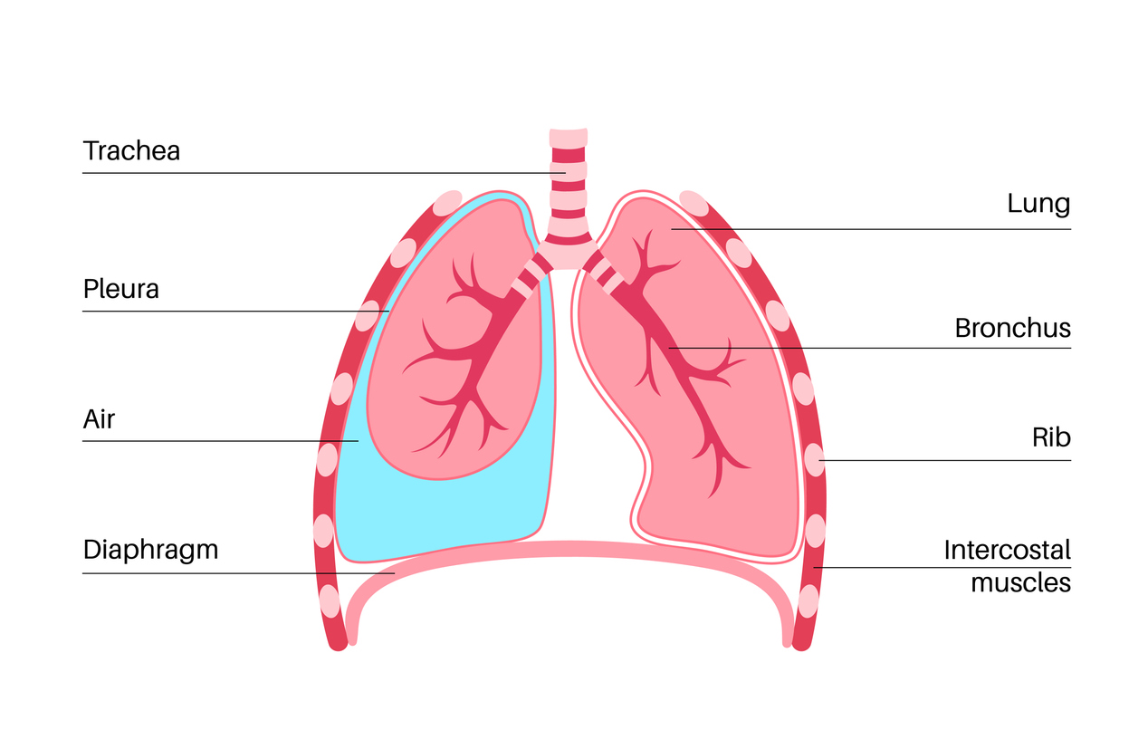
  • Visceral pleura (Pleura visceralis): The visceral pleura is the inner layer of the pleura and envelops both lungs. At the lung hilum (entry point of the pulmonary arteries, pulmonary veins, and bronchi into the lung), the visceral pleura transitions into the outer layer of the pleura (parietal pleura).
  • Parietal pleura (Pleura parietalis): The parietal pleura is the outer layer of the pleura and lines the chest wall (thoracic wall) and the upper surface of the diaphragm. Likely only the parietal pleura is innervated and is responsible for pain, for example in pleuritis.

Between the visceral pleura and the parietal pleura exists a sliding layer, because a pleural space (Cavitas pleuralis) is present. This space is filled with a few milliliters of serous fluid, the pleural fluid.

At the outer margins of the diaphragm and in the region of the mediastinum, the pleura forms so-called recesses (outpouchings), which serve as reserve folds during inspiration to allow expansion of the lung. A total of 4 recesses are distinguished:

  • Recessus costodiaphragmaticus
  • Recessus costomediastinalis
  • Recessus phrenicomediastinalis
  • Recessus vertebromediastinalis

The region of the pleura that extends superiorly (cranially) beyond the first rib is called the pleural cupula (Cupula pleurae).

Physiology of the Pleura:

The pleura serves as a sliding space between the chest wall and the lung. Through negative pressure in the pleural space (−5 cm H2O) and the capillary adhesion of the two pleural layers, the lung follows the movements of the chest wall and diaphragm, so that the musculature of these structures enables respiratory movements.

Pathophysiology of the Pleura:
When the negative pressure in the pleural space is eliminated by an injury (for example, pneumothorax), the lung can no longer follow the excursions of the respiratory muscles. With further pressure increase, collapse of the affected lung occurs.

In the pleural recesses, an abnormal amount of fluid (blood, exudate, pus, etc.) can accumulate. This is then referred to as pleural effusion (or hemothorax, pleural empyema).

Diseases of the Pleura
  • Accumulation of air in the pleural space: pneumothorax; occurring spontaneously or due to a traumatic event
  • catamenial pneumothorax in endometriosis;
  • Accumulation of air in the pleural space under pressure: tension pneumothorax;
  • Accumulation of blood in the pleural space: hemothorax; occurring after trauma or in pleural tumors;
  • Accumulation of fluid or pus in the pleural space in pleuritis;
  • Inflammation of the pleura (pleuritis); also in tuberculosis (TB);
  • Tumors of the pleura; pleural carcinomatosis (pleural metastases from another primary tumor); primary pleural tumors, especially malignant pleural mesothelioma (MPM). Benign solitary pleural fibroma.
14. January 2024 0 comments
FacebookLinkedinEmail
Thorax: Function and Anatomy

The Lungs (Pulmo)

by csadmin 14. January 2024
written by csadmin
Anatomy of the lungs:

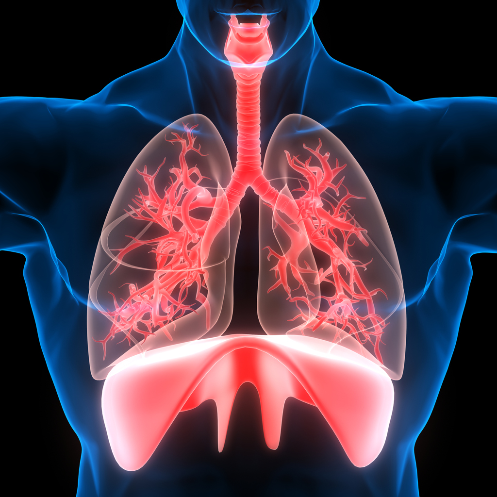
To ensure gas exchange, the lungs require close contact between the air circulation—maintained by breathing through the trachea and bronchi into the alveoli.

On the other hand, there must be close contact with the pulmonary arteries and pulmonary veins, which collectively ensure blood circulation down to the capillaries in the immediate vicinity of the alveoli.

In this way, venous blood in the pulmonary veins is cleared of carbon dioxide (CO₂) and enriched with oxygen (O₂). Like every organ, the lung is composed of segments, although physiologically well-defined boundaries are not always clearly demarcated anatomically.

Segmental pulmonary arteries, segmental pulmonary veins, and segmental bronchi circulate within these segments, enabling gas exchange in a very confined space. The segmental bronchus and segmental artery run together at the entry point of the lung segment (hilum), while the segmental vein runs separately.

Several lung segments are anatomically grouped into lung lobes. The right lung consists of three lobes: the upper lobe, the middle lobe, and the lower lobe. The left lung consists of only two lobes: the upper lobe and the lower lobe.
Histologically, the bronchi continue to divide via the respiratory bronchioles and alveolar bronchioles into the alveolar sacs, where gas exchange occurs.
Lung tissue serves gas exchange, the regulation of the acid-base balance, and has immunological and endocrine functions.

Diseases of the lungs (Pulmo):
  • Chronic obstructive pulmonary diseases (COPD, chronic bronchitis, smoker’s bronchitis); also in the context of chronic sinusitis
  • Gastroesophageal Reflux Disease
  • Bronchial asthma
  • Pulmonary fibrosis
  • Pulmonary arterial hypertension (high blood pressure in the pulmonary circulation)
  • Cystic fibrosis (CF, mucoviscidosis)
  • Pneumonia (pneumonias); also tuberculosis (TB)
  • Boeck’s disease (sarcoidosis)
  • Acute respiratory distress syndrome (ARDS)
  • Respiratory disorders in the context of other organ diseases (e.g., heart disease, brain disease, etc.)
  • Lung tumors, benign and malignant

Here you will find several expert interviews related to
lung diseases:

#08: Lung cancer diagnosis – what now?
Thorax-schweiz in conversation with Prof. Dr. med. Ralph Schmid

#05 Pulmonary nodules – what does the diagnosis mean?

Prof. Dr. Franzen spricht mit uns über Ursachen und Behandlung, Innovationen in der Diagnose und auch darüber, was Patient: innen selbst unternehmen können.

14. January 2024 0 comments
FacebookLinkedinEmail
Thorax: Function and Anatomy

The Airways (Trachea & Bronchi)

by csadmin 14. January 2024
written by csadmin
Anatomy of the Airways (Trachea, Bronchi):

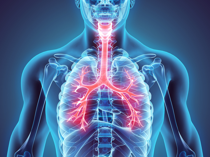
The respiratory tract is divided into the upper respiratory tract (nose, paranasal sinuses, and pharynx) and the lower respiratory tract (larynx, trachea, bronchi, and the lungs themselves). The airways in the narrower sense comprise the trachea and the bronchi. They transport air from outside to the alveoli during inhalation (inspiration) and back again during exhalation (expiration). Histologically, the bronchi have a single-layered ciliated epithelium with goblet cells. Typically, the bronchi have cartilaginous components in the wall that prevent the bronchus from collapsing during inhalation and exhalation.

Diseases of the Respiratory Tract (from the Nasal Cavity to the Alveoli):
  • Acute Respiratory Diseases (colds, rhinitis, acute sinusitis, acute otitis and pharyngitis, acute tonsillitis, common cold, influenza, acute bronchitis)
  • Allergic Lung Diseases
  • Alveolitis
  • Asbestosis
  • Bronchial Asthma
  • Chronic Sinusitis
  • Gastroesophageal Reflux Disease
  • COPD, Chronic Bronchitis and Pulmonary Emphysema
  • Pneumonia
  • Bronchiectasis
  • Tumors of the Respiratory Tract (malignant and benign) from the nose to the alveoli.
14. January 2024 0 comments
FacebookLinkedinEmail
Thorax: Function and Anatomy

Thorax: Function and Anatomy

by csadmin 14. January 2024
written by csadmin

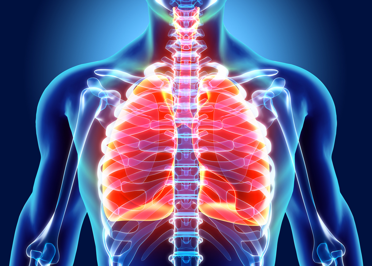
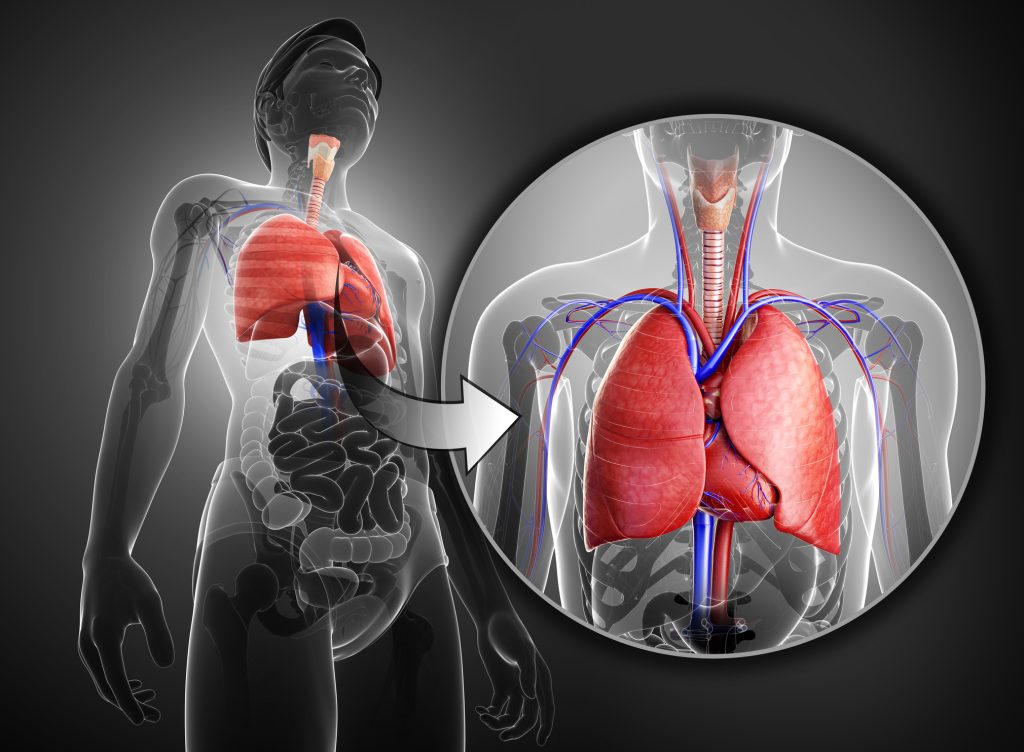
The thorax, also known as the chest, is a central region of the human body that encloses a variety of vital organs. This region extends from the upper part of the neck to the diaphragm and houses organs responsible for essential functions such as respiration and blood circulation.

The Anatomical Location of the Thorax:

The thorax is located between the neck (cervical spine) and the abdomen and consists of ribs, thoracic vertebrae, and the sternum. It forms a protective shell for vital organs and structures located in this area.

Which organs are located in the thoracic region?
  1. Heart: The heart is the engine of blood circulation and is located in the upper left part of the thorax.
  2. Lungs: The lungs occupy most of the thorax and are responsible for oxygen uptake and gas exchange.
  3. Bronchi and Trachea: These airways ensure the transport of air to and from the lungs.
  4. Thymus: The thymus is an essential organ of the immune system that plays a role in the development of T-lymphocytes.
  5. Esophagus: The esophagus enables the transport of food from the mouth to the stomach.
  6. Major Blood Vessels: The major blood vessels, such as the aorta and the vena cava, transport blood to and from various parts of the body.
Why do so many medical specialties focus on the thorax?

Medical professionals from various specialties, including cardiology, pulmonology, thoracic surgery, and radiology, are intensively dedicated to the study and treatment of thoracic diseases for several reasons:

  1. Vital Functions: The thorax houses organs that are crucial for vital functions such as respiration and blood circulation.
  2. Complexity and Interaction: The organs in the thorax work closely together, and problems in one area can affect others. A holistic approach is required to understand and address complex medical challenges.
  3. Diagnostics and Therapy: The diagnosis and treatment of diseases in the thorax require specialized knowledge and advanced technologies such as imaging techniques (e.g., X-ray, CT, MRI) and minimally invasive surgery.

Overall, the thorax forms a key area of the human body, laying the foundation for understanding and caring for vital life functions. The interdisciplinary collaboration of medical professionals from various specialties is crucial to ensure the health and well-being of patients.

14. January 2024 0 comments
FacebookLinkedinEmail
About Us

THE INITIATORS OF THORAX SWITZERLAND

by csadmin 13. January 2024
written by csadmin

Left: Tariq Abu-Naaj; right: Dr. med. Roland Kuster

Thorax Switzerland: together in search of clarity and support

The driving forces behind Thorax Switzerland are Dr. med. Roland Kuster and Tariq Abu-Naaj. With decades of experience in thoracic surgery, Dr. Kuster moved into medical management after a serious accident. Tariq Abu-Naaj, owner of Medical Brand, is involved in marketing for hospitals and medical professionals in Germany, Austria and Switzerland. He is particularly committed to ensuring that clinics take the perspective of patients into account in an exceptional and comprehensive way.

In a field that is unclear to many people—especially when it comes to thoracic medicine, where diagnoses are often serious and life-changing—the two have set themselves the goal of speaking with specialists across Switzerland in all areas of thoracic medicine. The aim: to create clarity and address the issues that truly matter to patients and their relatives.

Dr. med. Roland Kuster on the road for Thorax Switzerland

Thorax Switzerland is a long-term project that is expanded each month with additional contributions. Every question they receive is thoroughly examined by Dr. Kuster and Abu-Naaj in order to find satisfactory answers. They rely on blog posts as well as podcast interviews. The project is accompanied by a monthly newsletter that offers special topics for newsletter subscribers.

The Hirslanden Group, a private hospital group of Hirslanden AG in Switzerland with 17 clinics in 10 cantons and more than 3,000 attending physicians, supports the Thorax Switzerland project. This provides access to a unique network of resources and experts that leaves no questions unanswered. In addition, specialists from other clinics are warmly invited to participate in the project.

For Dr. Kuster and Abu-Naaj, the journey with Thorax Switzerland is a compelling search for answers to questions from patients and their relatives, in order to support them as effectively as possible. A journey they are looking forward to—one that helps shed more light on the often opaque field of thoracic medicine.

13. January 2024 0 comments
FacebookLinkedinEmail
About Us

Additional Experts

by csadmin 13. January 2024
written by csadmin

Additional experts expand the Thorax Switzerland network. In this section, we introduce an extended group of professionals who offer a broader perspective on thoracic medicine.

In addition to core topics such as lung cancer, COPD, sleep apnea, and asthma, further specialist areas are included. Experts from palliative care, spiritual care, oncological rehabilitation, as well as leaders of associations and self-help groups, share their knowledge and experience.

Our mission is to provide you not only with medical information but also with supportive resources for all aspects of your health journey.

We expand our network every month to ensure that you have access to the latest findings and a comprehensive range of expertise.

Discover the diverse world of thoracic medicine and be inspired by the valuable perspectives of our additional experts at Thorax Switzerland!

13. January 2024 0 comments
FacebookLinkedinEmail
About Us

The medical team at Thorax Schweiz

by csadmin 13. January 2024
written by csadmin

Thoracic medicine, a fascinating and multifaceted field, is comprehensively represented by the wide range of specialties within our medical team at Thorax Schweiz.

From lung cancer and COPD to sleep apnoea and asthma, our female experts cover a broad spectrum. Their different specialisations enable a holistic view of the challenges and solutions in the field of lung diseases. This diversity makes our medical team a valuable companion on your path to a healthy lifestyle. In addition, we expand our team of female experts every month to ensure that you always benefit from up-to-date and diverse medical expertise.

Discover the extensive expertise and support offered by our continuously growing group of specialist medical staff.

Prof. Dr. med. Othmar Schöb
Specialist in surgery, with a focus on thoracic and visceral surgery

Core competencies

Visceral surgery (surgery of the abdominal organs)

Major tumour surgery of the oesophagus, liver, pancreas, stomach, intestine, peritoneum and ovaries. Modern intraoperative cancer treatment of the abdominal cavity with hyperthermia and chemotherapy
(HIPEC and PIPAC)

Minimally invasive surgery and robot-assisted procedures

Gallstones
Diverticulitis
Colon cancer
Inguinal hernia
Hernia surgery
Reflux disease
Surgery for obesity (bariatric surgery)

Thoracic surgery (surgery of the lungs and thoracic organs)
Your contact for Prof. Dr. med. Othmar Schöb

Surgical Centre Zurich
Witellikerstrasse 40
8032 Zurich
CH
Telephone +41 44 387 37 00
www.chirurgischeszentrum.ch

Doctor direct
Telephone +41 44 387 29 66
Fax +41 44 387 29 69
Email os@professorschoeb.ch

Prof. Dr. med. Ulf Petrausch
Specialist in medical oncology, general internal medicine, allergology and clinical immunology

Core competencies

Oncology

Assessment and treatment of malignant neoplasms (tumours), in particular tumours of the prostate, kidney and urinary tract.

Immunology

Use and development of novel immunological tumour therapies

Thoracic surgery (surgery of the lungs and thoracic organs)

Assessment and treatment of diseases of the blood and immune system

Lymphomas

Including high-dose chemotherapy with autologous blood stem cell transplantation (ASCT) and CAR T-cell therapy

Your contact for Prof. Dr. med. Ulf Petrausch

OnkoZentrum Zurich
Seestrasse 259
8038 Zurich
CH
Telephone +41 43 344 33 33
Fax +41 43 344 33 44
Email enge@ozh.ch
www.ozh.ch

Doctor direct
Email ulf.petrausch@ozh.ch

Prof. Dr. med. Gregor Jan Kocher, MBA

Core competencies

Full spectrum of thoracic surgery with specialised expertise in minimally invasive surgical procedures, including robot-assisted techniques

Treatment of all intrathoracic pathologies, with the exception of cardiac surgical procedures

Minimally invasive procedures for lung tumours (e.g. lung cancer), tumours of the mediastinum (e.g. thymoma, cysts, schwannomas) and the pleura (malignant pleural mesothelioma, pleural effusion, pleural empyema, pneumothorax), as well as minimally invasive correction of chest wall deformities using a pectus bar (for funnel chest/pectus carinatum)

Surgical treatment of tracheal pathologies as well as thoracoscopic sympathectomy for erythrophobia and hyperhidrosis

Your contact for Prof. Dr. med. Gregor Jan Kocher

Bern Thoracic Surgery
Haus von Rodt
Schänzlihalde 1
3013 Bern
CH

Telephone +41 31 335 78 01
Fax +41 31 335 78 02
www.bernerthoraxchirurgie.ch

Doctor direct
gregor.kocher@hirslanden.ch
www.kocher-thoraxchirurgie.ch

Dr. med. Esther Bertschi
Specialist in medical oncology and general internal medicine

Core competencies

Treatment of all tumour diseases, outpatient and inpatient

Your contact for Dr. med. Esther Bertschi

Effingerstrasse 16
3008 Bern
CH
Telephone +41 31 301 89 26
Fax +41 31 301 89 27
www.onko-hirslanden-bern.ch

Doctor direct
esther.baertschi@hin.ch

Dr. med. Dr. phil. Jonas Klöpper
Specialist in medical oncology

Core competencies

Within medical oncology, I specialise in the treatment and follow-up care of patients diagnosed with a lung tumour, brain tumour or melanoma.

Your contact for Dr. med. Dr. phil. Jonas Klöpper

Clinic for Haematology & Oncology Hirslanden Zurich
Hirslanden Clinic
Witellikerstrasse 40
8032 Zurich
CH
Telephone +41 44 387 37 80
Fax +41 44 387 22 75
Email kho@hin.ch

Doctor direct
jonas.kloepper@kho.ch

13. January 2024 0 comments
FacebookLinkedinEmail
About Us

About Thorax Switzerland

by csadmin 13. January 2024
written by csadmin
This is how Thorax Switzerland helps patients navigate the complex world of thoracic medicine.
Find your way in thoracic medicine with Thorax Switzerland!

The world of thoracic medicine can be complex and confusing. Thorax Schweiz stands for expert advice and reliable information on thoracic diseases—born from the desire to provide comprehensive information. Today, Dr Roland Kuster and Tariq Abu-Naaj have made it their mission to provide people with thoracic diseases with comprehensive information.

The Thorax Schweiz concept was originally developed by Dr Roland Kuster and the late Faris Abu-Naaj. After Faris’ passing, Dr Kuster approached Faris’ brother, Tariq Abu-Naaj, to continue and expand this important initiative.

What goals are Dr med. Roland Kuster and Tariq Abu-Naaj, who lead the project, pursuing?

Together, we have set ourselves the goal of continuing the path begun by Roland and Faris and creating an information resource that competently guides patients and their relatives through the complex topics of thoracic medicine.

With this in mind, we are committed to travelling throughout Switzerland to meet physicians of all specialties in the field of thoracic diseases and to hold in-depth discussions with true experts in thoracic medicine.

The aim of Thorax Schweiz is to offer patients and their families solutions that truly help them.

Whether lung cancer, asthma, COPD, or sleep apnoea—we bring clarity to all aspects of thoracic medicine.
The well-being of patients is what drives us, and our approach is as sustainable as our mission: all our journeys are deliberately climate-neutral, by consistently using public transport.

Thorax Schweiz is not only an information platform, but also a community where everyone is welcome and invited to ask questions, expand their knowledge, and share it. We cordially invite medical specialists and lung specialists to join us and contribute their expertise.

Together, we are creating a network of knowledge and support that accompanies patients and their families on their journey.

Join the Thorax Schweiz community, too. We are here for you when you need help, information, and support.

13. January 2024 0 comments
FacebookLinkedinEmail
Newer Posts
Older Posts

Follow us for updates

Facebook Youtube Rss

Supported by

Supported by

Categories

  • About Us (4)
  • Blog (4)
  • Patients & Relatives (3)
  • Preventive Healthcare (5)
  • Thorax: Function and Anatomy (8)

About Us

About Us

helps patients navigate the complex world of thoracic medicine.

Points of Interest

  • Thorax: Function and Anatomy

  • The Pleura

  • The medical team at Thorax Schweiz

  • The Esophagus

Back Next

QuickLinks

  • Home
  • Contact
  • Imprint
  • Data Protection

@2024 - Copyright Hirslanden-Gruppe


Back To Top
Thorax Schweiz
  • Home
  • Anatomy & Disease Patterns
    • Thorax: Function and Anatomy
    • The Airways (Trachea & Bronchi)
    • The Lungs (Pulmo)
    • The Pleura
    • The diaphragm (diaphragma)
    • The mediastinum
    • The Chest (Thorax)
    • The Esophagus
  • Patients & Relatives
    • Patients & Relatives
    • Support Groups and Organizations
    • Your General Practitioner’s Office
  • Prevention
    • Preventive Healthcare
    • The Importance of Prevention
    • Smoke-Free Living
    • Follow-up care
    • Rehabilitation
  • Podcast
  • Blog
  • About Us
    • About Thorax Switzerland
    • The medical team at Thorax Schweiz
    • Additional Experts
    • THE INITIATORS OF THORAX SWITZERLAND
  • Contact
  • English
    • Français (French)
    • Deutsch (German)
    • Italiano (Italian)
  • English
  • Français (French)
  • Deutsch (German)
  • Italiano (Italian)